Ultrasound Examination
Ultrasound Examination
A non-invasive and safe diagnostic method that uses sound waves to visualize internal organs and tissues. Sound waves reflect from internal organs and create an image on the monitor, which enables physicians to precisely assess fetal development and identify possible abnormalities.
- Ultrasound examination is safe also during pregnancy
- It can be performed repeatedly without concerns for the health of the mother and fetus.
In our center we have specialized staff and modern equipment for reliable diagnostics.
PERFORMED ONLY AT GYN-FIV BRATISLAVA CENTER.
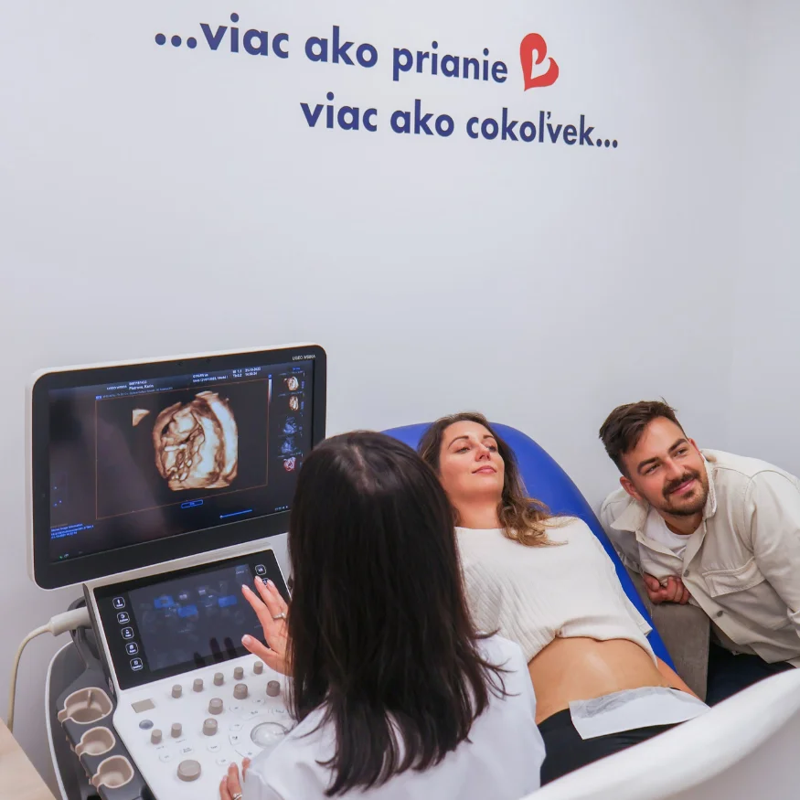
Benefits of Ultrasound Examination During Pregnancy
It helps detect possible abnormalities in the mother and fetus, as well as monitor the overall development of the pregnancy.
The main benefits include:
- determining gestational age
- identifying abnormal accessory organs and problems with the uterus
- assessing fetal growth and development
- checking fetal organ development
- evaluating placenta condition
- detecting genetic abnormalities of the fetus
- monitoring amniotic fluid volume
- detecting multiple pregnancies
Scheduling an Examination
To schedule an ultrasound gynecological examination during pregnancy, it is necessary to bring a referral from your primary care gynecologist.Screening Ultrasounds at Important Stages:
I. Screening Examination (Week 11 - 14 of Pregnancy)
It serves to confirm the number of fetuses, determine the length of pregnancy and rule out ectopic pregnancy. It also helps in diagnosing certain genetic risks and checks fetal viability, including monitoring of cardiac activity.
II. Fetal Morphological Examination = Morphological Ultrasound (Week 20 - 22 of Pregnancy)
Also called the detailed ultrasound.
The physician examines: the shape and number of limbs and internal organs, possible deviations from the norm and their severity; the number of fetuses – similar to the first trimester, checking whether you are expecting one or more children; checks the shape, position and morphology of the heart; measures the fetus, approximately determining weight and size; checks the length of the cervix and placenta position; may determine the baby's sex – though this is not always possible, sometimes the baby simply "hides".
III. Screening Examination (Week 29 - 32 of Pregnancy)
It monitors fetal growth and development, amniotic fluid volume, baby's position, placenta condition and blood flow in the umbilical cord. The examination helps verify that the pregnancy is progressing normally in the final trimester as well.
IV. Screening Examination (Week 36 of Pregnancy)
The physician checks fetal position, placenta condition, amniotic fluid volume and prepares the expectant mother for delivery. It also monitors the possible presence of risk factors that could affect the method of delivery management.